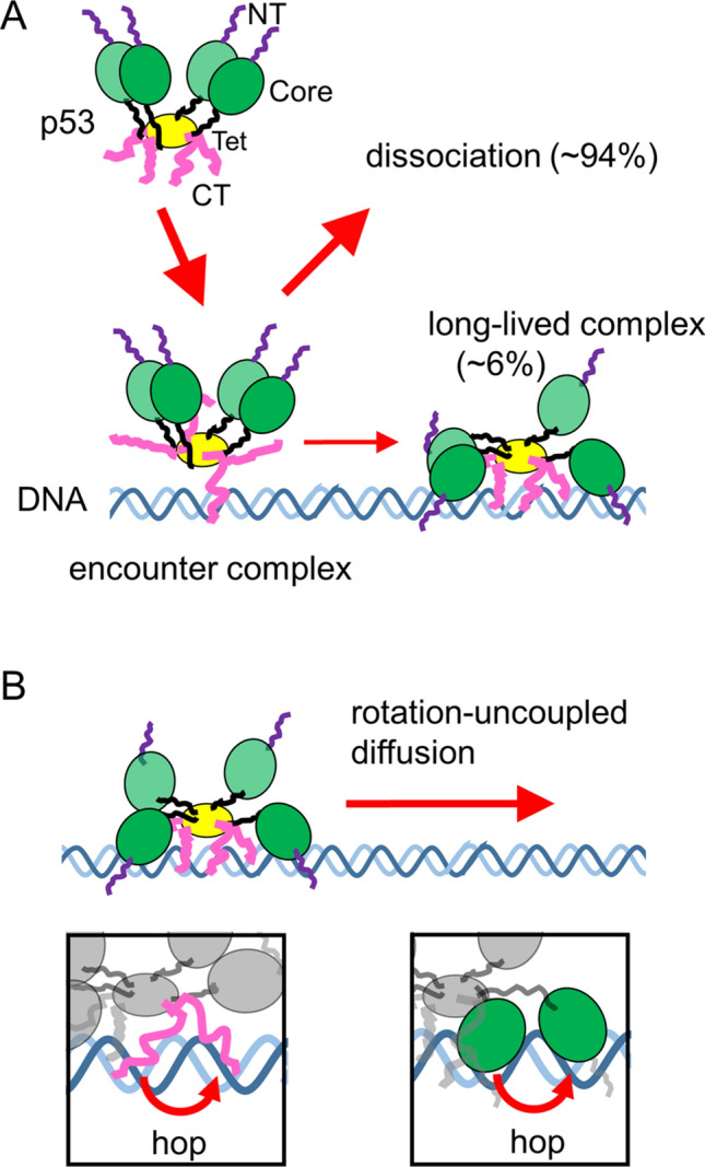
Figure 6.

Proposed model of the target search dynamics of p53 based on sub-millisecond-resolved single-molecule measurements. (A) Schematic diagram of the encounter and long-lived complexes of p53 tetramer and DNA. NT, Core, Tet, and CT respectively denote the N-terminal, core, tetramerization, and C-terminal domains of p53. At least one of the CT domains interacts with DNA in the encounter complex, and other DNA-binding domains are recruited to form the long-lived complex in the subsequent step. (B) Schematic diagram of the rotation-uncoupled motion of p53 in the presence of physiological salt concentrations. The CT and/or core domains hop from one phosphate backbone to the other backbone separated by a half turn of the helix, resulting in rotation-uncoupled diffusion (insets).
