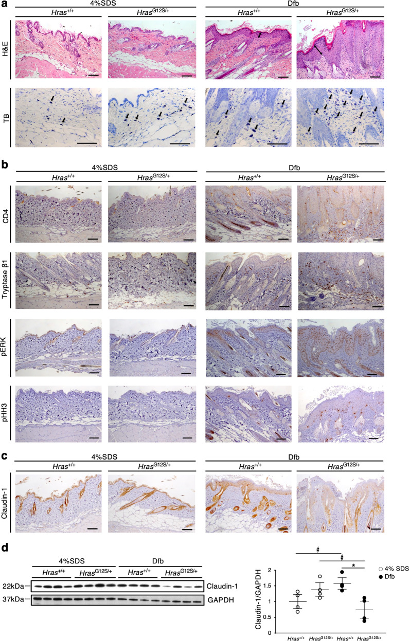
Fig. 2. Histological analysis reveals acanthosis with hyperproliferation of p-ERK-positive epidermal cells, increased inflammatory cells, and reduced claudin-1 expression in the dorsal skin of Dfb-treated HrasG12S/+ mice.
a Skin tissue stained with H&E and TB. b, c Immunohistochemistry of CD4, tryptase β1, p-ERK, pHH3, and claudin-1 in the skin. a–c Scale bars: 100 μm. d Lysates from the skin were immunoblotted with anti-Claudin-1 antibody. Band intensities were quantified and compared among the four groups. The expression levels were normalized to GAPDH (n = 4 in each group). Data are presented as mean ± SD. Significance was analyzed by one-way ANOVA and the Tukey−Kramer method. *P < 0.05, #P < 0.05, two-tailed Student’s t test.

