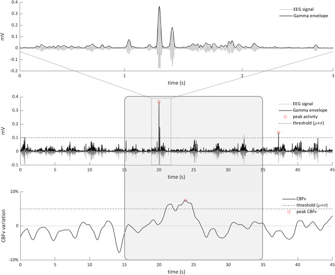
Fig. 1.
Schematic of analysis techniques used to identify neural events for selecting epochs of the accompanying hemodynamics. Magnification of the gamma filtered EEG signal and the corresponding envelope (Eγ) of a peak of neuronal activity (top row). A 45 s Eγ trial from a single animal is shown as an example (middle row); two peaks of neuronal activity above the chosen threshold are marked. A time epoch of 20 s around the first peak is drawn. Corresponding 45 s of CBFv fluctuation from the same animal (bottom row); an increase in the CBFv appears few seconds after the Eγ peak

