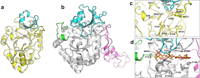
Figure 2.
Structure of FjChiB. The overall structure of FjChiB (a) is shown next to SmChiB (b; PDB accession 1e6n). The chitinase insertion domain (CID) of each protein is colored cyan, and the -3-site capping loop and CBM family 5 domain of SmChiB are colored green and magenta, respectively. The active site architectures of FjChiB (c) and SmChiB (d) show the key residues lining the substrate binding clefts. The structure of SmChiB contained the catalytic residue substitution E144Q enabling the determination in complex with chitopentaose (orange sticks). Notably, the smaller CID and the lack of motifs equivalent to the capping loop of SmChiB lead to a more exposed substrate binding cleft in FjChiB.

