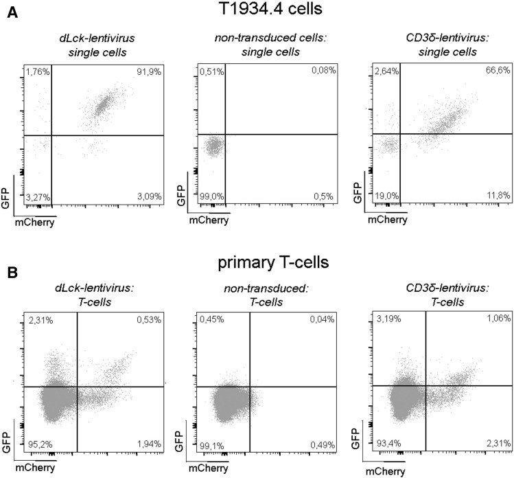
Figure 2.
Functionality of bicistronic lentiviral constructs in vitro. (A) T1934.4 cells were transduced using MOI 50 with the dLck-lentivirus or the CD3δ-lentivirus. eGFP and mCherry expression were analyzed 7 days post lentiviral transduction. Transduced cells (right and left) are double positive for eGFP and mCherry compared to non-transduced cells (middle). (B) Primary murine spleen T-cells were infected with the respective lentivirus as depicted and expanded in vitro using CD3xCD28 costimulation for 3 days in a single transduction experiments. The dot plots depict eGFP and mCherry expression for dLck-lentivirus (middle) and CD3δ-virus (right) treated T-cells gated on mCherry positive and negative subpopulations.

