Figure 4.
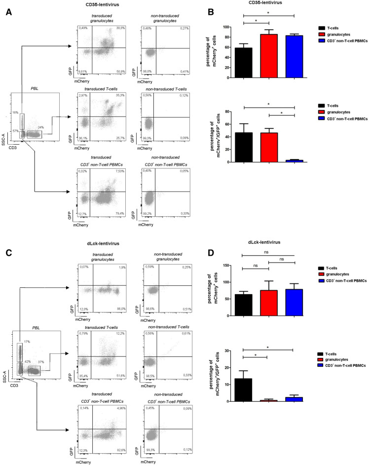
Specificity of lentiviral constructs in peripheral blood. Eight to ten weeks post HSC-transplantation leukocyte subsets in peripheral blood were evaluated by flow cytometric analysis for CD3δ-lentivirus transduced HSCs (n = 5, A,B) or for dLck-lentivirus transduced HSCs (n = 9, C,D). Representative dot plots depicting eGFP and mCherry expression are shown for CD3+ T-cells (A,C, left), CD3− non-T-cell PBMCs (A,C, middle) and CD3− granulocytes (A,C, right). In (B,D) quantification of mCherry+ and GFP+ cells. Error bars indicating SD. *p < 0.05.

