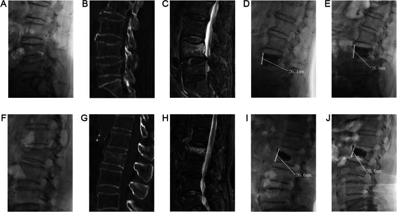
Fig. 2.
Typical cases. A 71 years old female patient, preoperative x-ray (a), CT (b), MRI (c) showed acute OVCF of L4. X-ray (d) at 24 h post operation showed that the cement was in close contact with the upper and lower endplates, and X-ray (e) at 12 months post operation showed that the vertebral height was maintained well. A 62 years old female patient, preoperative x-ray (f), CT (g), MRI (h) showed acute OVCF of L2. The X-ray (i) at 24 h post operation showed that the cement did not contact the lower endplate. The X-ray (j) at 6 months post operation showed that the height of the vertebral body was lost and the vertebral body was recompressed

