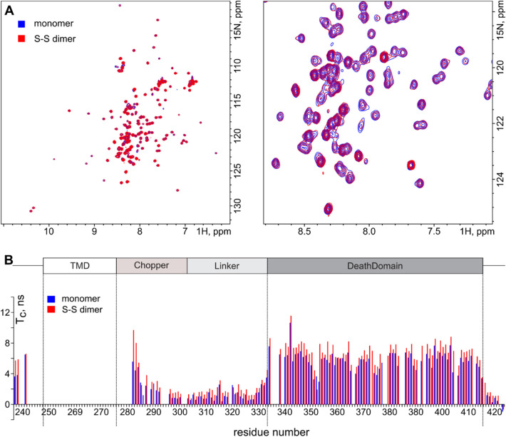
Figure 5.
The effect of dimerization on the structure and behavior of the intracellular domain of rP75-ΔECD-3CX. (A) 1H,15N-HSQC spectra of rP75-ΔECD-3CX monomer (shown in blue) and disulfide-crosslinked dimer (shown in red). The full spectrum and central region are shown at left and right, respectively. (B) NMR-derived rotational diffusion correlation time of N–H bonds is plotted versus the residue number of rP75-ΔECD-3CX monomer (blue bars) and dimer (red bars). The figure was prepared using the program Inkscape 0.92 (https://inkscape.org/).

