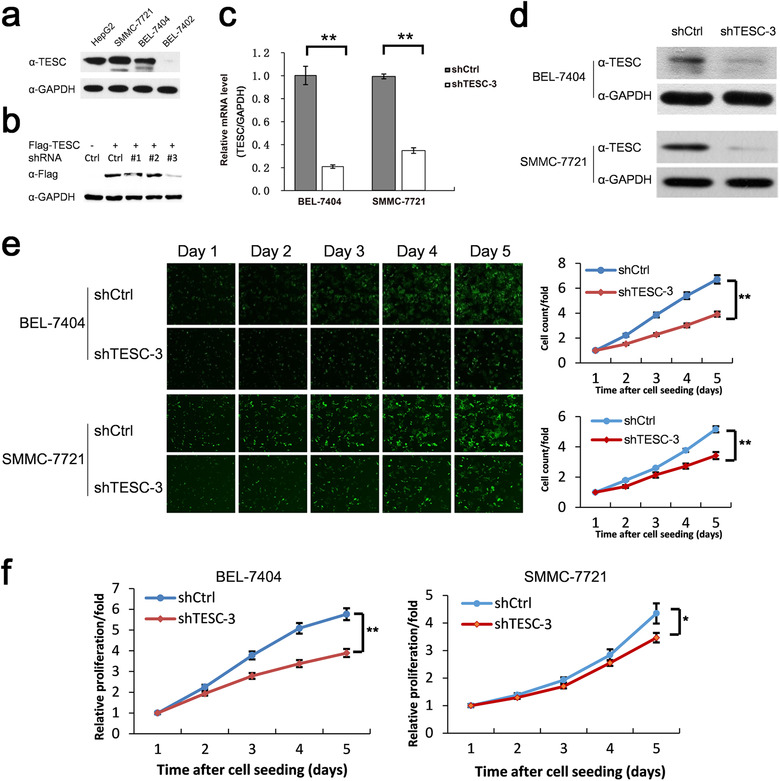
FIGURE 3.

Association of TESC with HCC cell growth. a. Western blotting analysis of TESC expression in different liver cancer cell lines. b. Western blotting analysis of cell lysates of HEK293T transfected with Flag‐tagged TESC and 3 pairs of TESC shRNA (#1‐#3) or the control shRNA (Ctrl) after 36 hours with indicated antibody. c, d. RT‐qPCR (c) and Western blotting (d) analyses of BEL‐7404 and SMMC‐7721 cells infected with lentiviruses containing TESC shRNA (shTESC‐3) or the control shRNA (shCtrl). e. Celigo cell counting of BEL‐7404 and SMMC‐7721 cells infected with lentiviruses containing shTESC‐3 or shCtrl. f. WST‐1 assay of BEL‐7404 and SMMC‐7721 cells infected with lentiviruses containing shTESC‐3 or shCtrl. *: P < 0.050; **: P < 0.010. TESC: Tescalcin; HCC: hepatocellular carcinoma; shRNA: short‐hairpin RNA; RT‐qPCR: real‐time quantitative polymerase chain reaction
