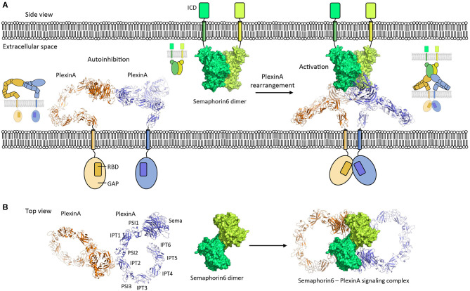
Figure 5.
PlexinA autoinhibition model and activation by Semaphorin6 ligand. (A) PlexinA receptors adopt an autoinhibited state by non-symmetric cis dimerization (pdb 5l5k). The transmembrane helixes are separated from each other in this state. Rearrangement of the PlexinA dimer upon Semaphorin6 ligand binding (pdb 3okw) activates the PlexinA receptors by bringing the transmembrane helixes in close proximity (modeled based on 3oky and 5l5k). ICD, intracellular domain; GAP, GTPase activating protein domain; RBD, Rho GTPase binding domain. (B) Top view [i.e., (A) is rotated by 90° along the membrane]. The membranes and cytosolic segments are omitted from the panel. PSI, plexin-semaphorin-integrin; IPT, Ig domain shared by plexins and transcription factors.

