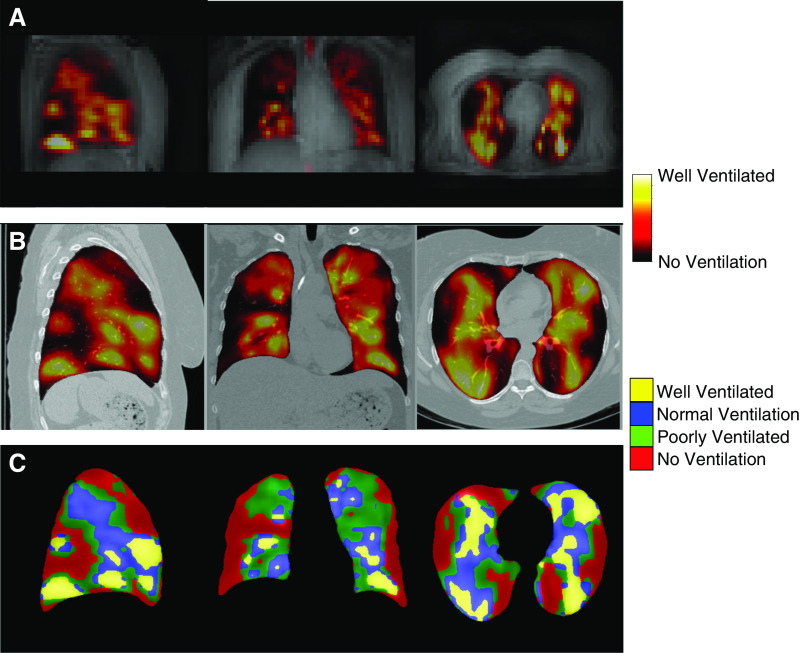
Figure 2.
129Xe magnetic resonance imaging (MRI) images in severe asthma. (A) Low-resolution proton MRI with ventilation image overlay of a patient with severe asthma prior to bronchial thermoplasty. (B) High-resolution volumetric computed tomography with MRI ventilation image overlay after registration to the computed tomography space. (C) The continuous ventilation image is segmented into four categories (no ventilation, poor ventilation, normal ventilation, and well ventilated).

