
Fig. 1. Crystal structure of human AdipoR1(A208).
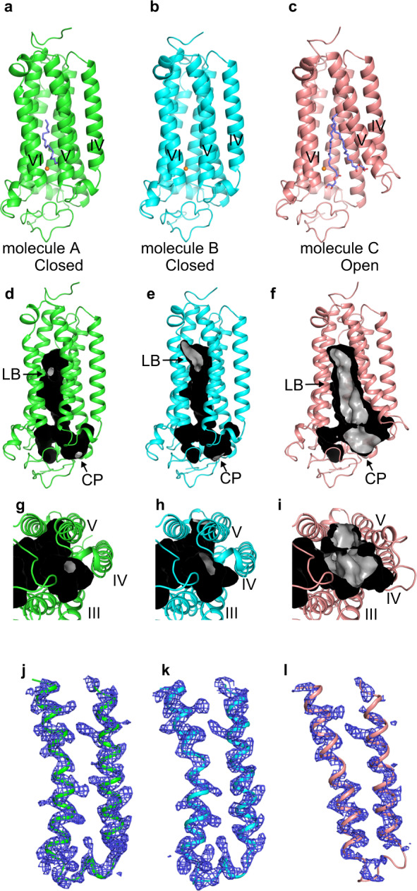
The crystal structure of human AdipoR1(A208) at 3.1-Å resolution: the asymmetric unit contains three molecules, A, B, and C (a–c). Molecules A (green) (a) and B (cyan) (b) are in the closed form, and molecule C (salmon) (c) is in the open form. The structures are viewed parallel to the membrane. The zinc ion is shown as an orange sphere. Two oleic acid molecules in molecule A and two monoolein molecules in molecule C are shown as slate blue stick models. d–i The major cavities of molecules A (d, g, green) and B (e, h, cyan) in the closed form, and molecule C (f, i, salmon) in the open form of AdipoR1(A208). The outside and inside surfaces of the major cavities are colored black and gray, respectively. The major cavities are viewed parallel to the membrane (d–f) and from the intracellular side (g–i). The lipid-bilayer (LB) and cytoplasmic (CP) openings are labeled LB and CP, respectively. Other minor cavities are omitted for clarity. j–l The simulated-annealing Fo–Fc omit maps, contoured at 2.5σ. The electron densities on helices IV and V and ICL2 of molecules A (j, green) and B (k, cyan) in the closed form, and molecule C (l, salmon) in the open form of AdipoR1(A208). For comparison, molecules C and A are shown in gray in j/k and l, respectively.