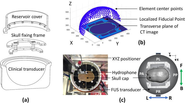
Fig. 6.
The registration of clinical focused ultrasound system and cadaver skull. a Computer-aided design (CAD) sketch of the clinical transducer, skull fixing frame, and reservoir cover. b Fiducial point-based registration of the transducer elements and skull volume in scanned CT images. Notably, the detailed geometry of the actual FUS transducer was excluded in this study. The sketch in (a) and (b) does not represent the actual scale of the FUS transducer. c Top view illustrating the coordinate configuration, including patient perspective coordinate (patient’s anterior-PA & patient’s posterior-PP, patient’s left-PL & patient’s right-PR), CT image coordinate, and coordinate of hydrophone scanning system (front [F] & back [B], left [L] & right [R])

