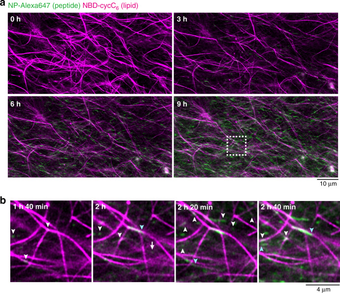
Fig. 6. Real-time imaging of formation process of the parallel self-sorting network.
a Time-lapse imaging of the formation process of the parallel SDN. b Magnified view of seed formation on the surface of lipid-type nanofibers (a white square in Fig. 6a). White and sky blue arrowheads represent the sites of seed formation and elongation on the surface of lipid nanofibers, respectively. Green: NP-Alexa647, magenta: NBD-cycC6. Condition: [Ald-F(F)F] = 17.3 mM (0.80 wt%), [Phos-MecycC5] = 2.4 mM (0.15 wt%), [carboxymethoxylamine] = 20.8 mM (1.2 eq), [O-benzylhydroxylamine] = 69.2 mM (4.0 eq), [NP-Alexa647] = 4.0 µM, [NBD-cycC6] = 4.0 µM, 100 mM MES, pH 6.0, 30 °C.

