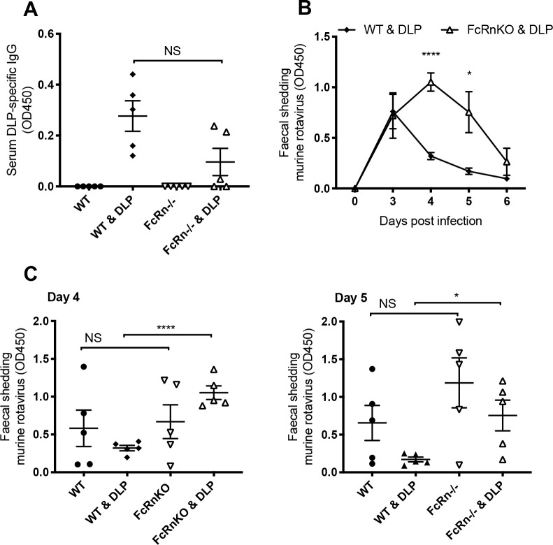
Fig 5. Rotavirus infection in FcRn deficient mice.
(A) DLP-specific antibodies in serum of naïve and DLP-immunized, WT and FcRnKO mice (5 mice per group, each symbol representing one mouse). (B) Faecal antigen shedding on days 0–6 post infection as detected by ELISA in the mice pre-immunized with DLPs, comparing WT with FcRn knockout (FcRnKO) mice. (C) Amounts of EMcN virus shedding in faeces of the naïve and DLP-immunized, WT and FcRnKO mice, days 4 and 5 of experiment shown in panel (B). Horizontal lines represent mean and standard error, **** p = < 0.0001, * p = < 0.05, NS not significant.

