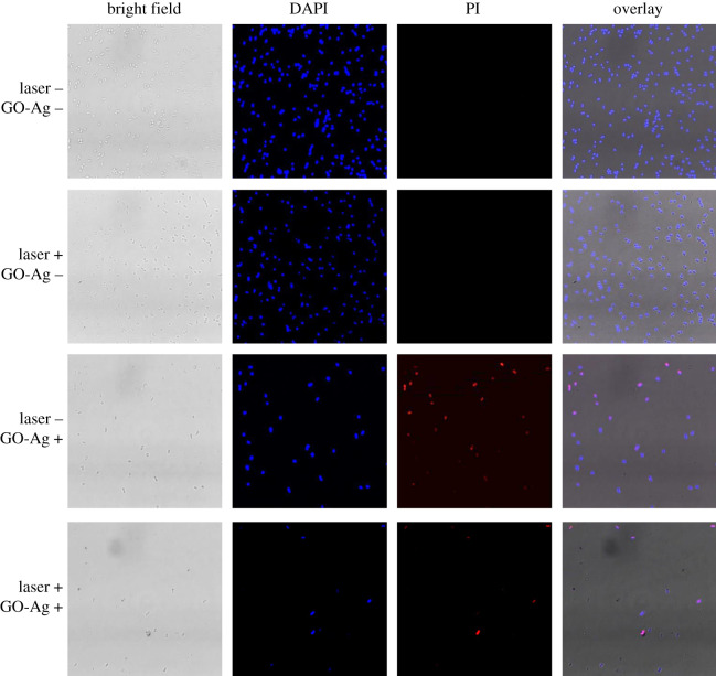
Figure 4.
Fluorescence imaging of MDR-2 E. coli with and without photothermal treatment. DAPI and PI were used to stain the nuclei acid of bacteria. The difference is DAPI stains all the bacteria while PI can only stain the non-living bacteria. DAPI-stained bacteria emit blue fluorescence light while PI-stained bacteria emit red fluorescence light. ‘Laser +’ and ‘Laser –’ denote the presence and absence of photothermal treatment, ‘GO-Ag +’ and ‘GO-Ag –’ denote the presence and absence of GO-Ag nanocomposites. 1.5 W cm−2, 7 µg ml−1 GO-Ag. Scale bar is 5 µm.

