
Fig. 1. MIZ1 protein levels are reduced in human COPD lungs and cigarette smoke–exposed mouse lungs or cigarette smoke extract–treated lung epithelial cells.
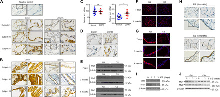
(A and B) Miz1 immunohistochemistry (IHC) staining of normal human lungs or lungs from patients with COPD at the time of lung transplantation (three subjects from each group are shown, which were representative of seven subjects from each group). Scale bars, 20 μm. (C) mRNA levels of Miz1 and Mule from normal human lungs or lungs from patients with COPD analyzed by RNA sequencing (RNA-seq) (normal, n = 13; COPD, n = 7). **P < 0.01; ns, not significant. (D) Mule IHC from human lungs mentioned in (A). (E to H) Western blot (E) or IF staining (F and G) of Miz1 or Mule IHC (H) in mouse lungs exposed to RA or CS for 7 days, 3 months, and 6 months. Representatives of n ≥ 4. (I and J) Western blot of Mule and Miz1 in CS extract–treated MLE-12 cells (I) or cells expressing tetracycline-inducible shRNA for Mule in the absence or presence of tetracycline (Tet) (J). Data are representative of at least three independent experiments.