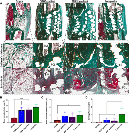
Fig. 6. Spatiotemporal delivery of both VEGF and BMP-2 led to enhanced bone healing and tissue morphology 12 weeks after scaffold implantation.

(A) Goldner’s trichrome– and Safranin O–stained sections of all groups after 12 weeks in vivo. Images were taken at 20×. BM denotes bone marrow. PCL denotes areas where the PCL frame was. DB denotes cartilage undergoing endochondral ossification to become new bone, and B denotes positive bone tissue. Quantification of the amount of (B) bone formation, (C) bone marrow, and (D) developing bone per total area. Error bars denote SDs. *P < 0.05, **P < 0.01, and ****P < 0.0001 (n = 9 animals).
