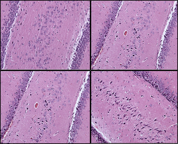
FIGURE 5.

Hippocampus (CA1‐4 region) ischemic and healthy neurons presentation (HE, ×200). Images were taken at 24 hr (upper) and 72 hr (lower) of reperfusion after administration of medication. BPC 157 (left) or saline (control, right) at 24 hr (upper) and 72 hr (lower) of reperfusion after surgery
