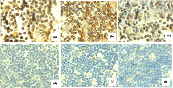
FIGURE 1.

Immunohistochemical staining for hypoxia‐inducible factor 1α (HIF‐1α), glucose transporter 1 (GLUT1), and hexokinase 2 (HK2). (a‐c) A primary central nervous system lymphoma sample that was positive for HIF‐1α, GLUT1, and HK2. (d–f) A normal lymph node sample that was negative for HIF‐1α, GLUT1, and HK2
