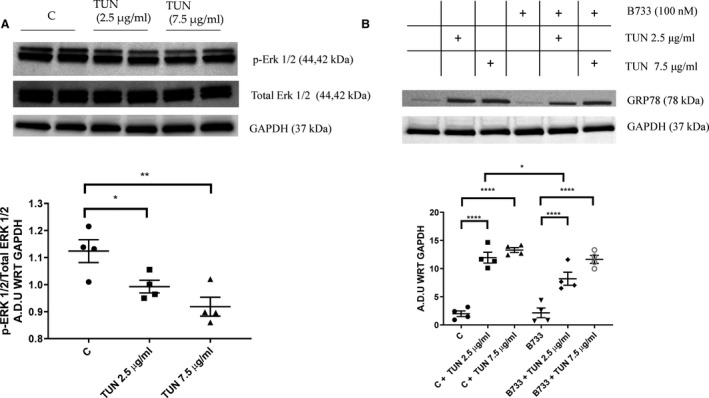
Figure 7. Western blot analysis of tunicamycin‐induced endoplasmic reticulum (ER) stress in protein lysates from primary cardiomyocytes.

Exposure of primary cardiomyocytes to tunicamycin (TUN) for 24 hours significantly reduces phosphorylation of extracellular signal‐regulated kinase (Erk) 1/2 (p‐Erk 1/2) at both 2.5 and 7.5 μg/mL concentrations of TUN (A); and significantly increases expression of GRP78 at both doses of TUN (B). B7‐33 (100 nmol/L) significantly lowers GRP78, compared with control (C), on coincubation with 2.5 μg/mL TUN (for A and B, n=4–6 experiments/group). Western blots are quantified and expressed as arbitrary densitometric units with respect to (A.D.U WRT) the expression of GAPDH. For A and B, 1‐way ANOVA was used for significance testing, and if P<0.05, Holm‐Sidak test was used for post hoc analysis. *P<0.05, **P<0.01, ****P<0.0001.
