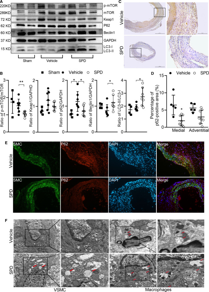
Figure 6. Spermidine treatment increases autophagy‐related proteins in experimental AAAs.

Aortae were collected 14 days after PPE infusion. Western blot, immunohistochemical staining and transmission electron microscopic examination were performed to evaluate the autophagy in experimental AAA. Protein levels of p‐mTOR, mTOR, Keap1, p62, Beclin1, GAPDH, LC3‐I, and LC3‐II were detected by Western blot (A) and quantified by densitometry (B). Data are presented as mean±SD. Unpaired t test, **P<0.01 and *P<0.05 vs vehicle group, n=4 to 7 in each group. C, Representative immunostaining images for p62 protein in vehicle (n=5) and SPD (n=8) groups. Scale bar=50 μm, 100 μm. D, p62 protein in medial (P=0.001) and adventitial (P=0.054) segments of aneurysm was quantified as p62‐positive area. Two‐way ANOVA, *P<0.05 vs vehicle group. E, Representative immunofluorescence staining with antibodies against p62 protein conjugated with Alexa Fluor 594 (red color), and antibodies against SMA conjugated with Alexa Fluor 488 (green color), with the nucleus counterstained with DAPI (blue color). F, Representative transmission electron microscopy of aorta in two groups. AAA indicates abdominal aortic aneurysm; DAPI, 4′,6‐diamidino‐2‐phenylindole; GAPDH, glyceraldehyde‐3‐phosphate dehydrogenase; Keap1, kelch‐like ECH associated protein 1; LC3, microtubule‐associated protein 1 light chain 3; mTOR, mammalian target of rapamycin; PPE, porcine pancreatic elastase; and SPD, spermidine.
