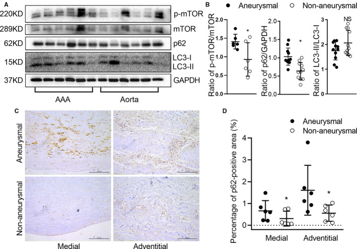
Figure 7. Evidence for dysregulated autophagy in clinical AAAs.

Western blot and immunostaining were performed to evaluate the autophagy of AAA and adjacent normal aortas from patients who underwent open AAA repair surgery. Protein levels of p‐mTOR, mTOR, p62, LC3‐I, and LC3‐II were detected by Western blot (A) and quantified by densitometry (B). Data are presented as mean±SD. Paired t test, NS for nonsignificance, *P<0.05 vs AAA, n=6 to 11. C, Representative aortic histology images of immunohistochemical staining of p62 protein. D, Analysis of p62 protein in medial (P=0.025) and adventitial (P=0.036) in adjacent normal and aneurysmal human aortae. Scale bar=100 μm. Data are presented as mean±SD. Paired t test, *P<0.05 vs AAA, n=6 in each group. AAA indicates abdominal aortic aneurysm; LC3, microtubule‐associated protein 1 light chain 3; and mTOR, mammalian target of rapamycin.
