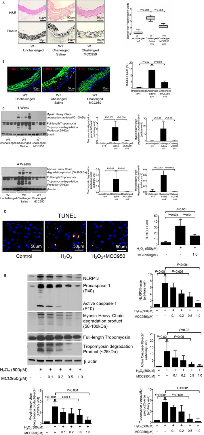
Figure 2.

Attenuation of challenge‐induced aortic degeneration, elastic fiber fragmentation, and reduced smooth muscle cell (SMC) apoptosis and SMC contractile protein degradation in vitro and in vivo after MCC950 treatment. A, Hematoxylin and eosin (H&E) staining and Verhoeff–van Gieson elastin staining showing that challenge‐induced aortic degeneration and elastic fiber fragmentation were reduced in mice treated with MCC950 after 4 weeks of angiotensin II (AngII) infusion. Elastic fiber fragmentation grades are also shown. B, Representative terminal deoxynucleotidyl transferase dUTP nick‐end labeling (TUNEL) staining showing that MCC950 prevented SMC apoptosis in challenged wild‐type (WT) mice. C, Representative Western blot images and quantification showing that MCC950 decreased levels of myosin heavy chain and tropomyosin degradation in challenged WT mice after 1 week and 4 weeks of AngII infusion. SMCs were treated with increasing doses of MCC950 as indicated. D, Representative TUNEL staining showing that MCC950 prevented SMC apoptosis induced by hydrogen peroxide (H2O2). E, Representative Western blot images and quantification showing that MCC950 dose‐dependently decreased the levels of nucleotide‐binding oligomerization domain–like receptor pyrin domain containing 3 (NLRP3), caspase‐1, myosin heavy chain, and tropomyosin degradation. Data are presented as mean±SEM. One‐way ANOVA with the post hoc Dunnett T3 test was used for pairwise comparisons in (A, B, and D). The post hoc Tukey test was used for pairwise comparisons in (C and E). DAPI indicates 4′,6‐diamidino‐2‐phenylindole.
