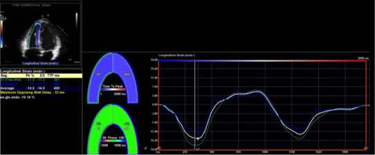
Figure 2. Representative image of right ventricular strain measurement.

A sample image from TomTec Image‐Arena is shown. Software detected the myocardial–endocardial interface and was manually adjusted by an operator to ensure accuracy. The software then calculated strain and strain rate.
