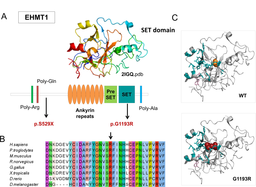
Figure 3. EHMT1 missense mutation affects SET domain.

Causative variants identified in our patient cohort are mapped to the EHMT1. Protein sequence presents regions biased toward polar (glutamine and arginine) and a poly-alanine motif. The ankyrin domain (orange) is involved with the histone H3K9me binding. The Pre-SET domain (green) contributes to SET domain stabilization B) EHMT1 p.Gly1193 and neighboring residues are conserved among orthologous sequences. Amino acids are colored by conservation, according ClustalX color code. C) EHMT1 p.Gly1193Arg variant (red) and wild type glycine (orange) are mapped to SET domain structure (2igq.pdb, chain A) Residues involved in H3K9 binding and the S-adenosyl-L-methionine molecule are represented in blue sticks.
