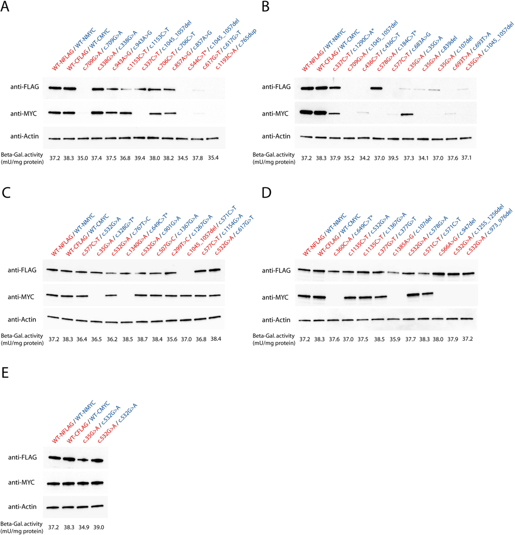
Figure 2. Overview of protein expression levels per variant combination.
COS-7 cells were transfected with 2.5 μg of each FLAG-and MYC-tagged ASL expression vectors and 1 μg of β-galactosidase reporter plasmid, cultured for 48 hours and subjected to protein expression analysis using standard Western blot technique. Briefly, proteins were separated by sodium dodecyl sulfate polyacrylamide gel electrophoresis (SDS-PAGE) and blotted on nitrocellulose membranes using the Trans-Blot® Turbo Transfer System (BioRad). Expression of FLAG-or MYC-tagged ASL variants was visualized using anti-FLAG-or anti-MYC antibodies on two identical gels, which were carried in parallel (A-E). Equal protein loading in cell lysates was confirmed by immunoblotting using an anti-β-actin antibody. Activity of β-galactosidase per variant combination was measured applying the β-galactosidase enzyme assay system (Promega). Data are expressed as mean in mU/mg protein (A-E, n=3). Of note, full-size images of anti-FLAG and anti-MYC stainings per variant combination are depicted in Supp. Figure S1. Variants associated with a premature stop codon are indicated by an asterisk (*). Red letters illustrate FLAG-tagged expression vectors, blue letters represent MYC-tagged plasmids.

