
Figure 4. Effects of HCQ and QC on NF-κB activation.
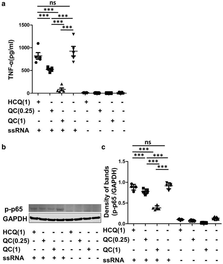
Peripheral blood mononuclear cells isolated from five cutaneous lupus patients were treated with increasing concentrations of QC (0.25–1 μg/ml) and HCQ (1 μg/ml) for 3 hours, with or without ssRNA stimulation. (a) TNF-α level in the culture medium after 3 hours was evaluated by ELISA. (b) Representative band from Western blot analysis for activated NF-κB (phospho-p65). GAPDH was used as the loading control. (c) The density of each band was analyzed with ImageJ software (National Institutes of Health, Bethesda, MD) and presented as fold changes normalized to GAPDH. p-p65, phospho-p65. The graphs show mean ± SEM (n = 5). GAPDH, glyceraldehyde-3-phosphate dehydrogenase; HCQ, hydroxychloroquine; QC, quinacrine; ssRNA, single-stranded RNA. ***P < 0.001.