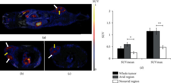
Figure 3.

In vivo PET/MRI studies of αvβ3 integrin receptor expression in subcutaneously transplanted He/De tumors. Representative coronal (a) and axial (b, c) PET/MRI images of subcutaneously transplanted He/De tumors 13 days after tumor induction and 90 min after intravenous injection of 68Ga-NODAGA-[c(RGD)]2. White arrows: αvβ3 integrin receptor positive (avid) regions; yellow arrows: nonavid regions. (d) Quantitative PET/MRI image analysis of heterogenous He/De (n = 15) tumors. Significance levels: p ≤ 0.05 (∗) and p ≤ 0.01 (∗∗). Data is presented as the mean ± SD.
