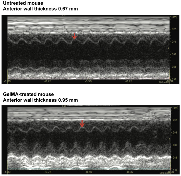
Figure 4. Transthoracic echocardiography after myocardial infarction.

M‐mode echocardiograms in representative untreated (top) and gelatin methacryloyl (GelMA)–treated (bottom) mouse hearts. Measurement of anterior left ventricular wall thickness denoted by the location of the red arrow in each panel.
