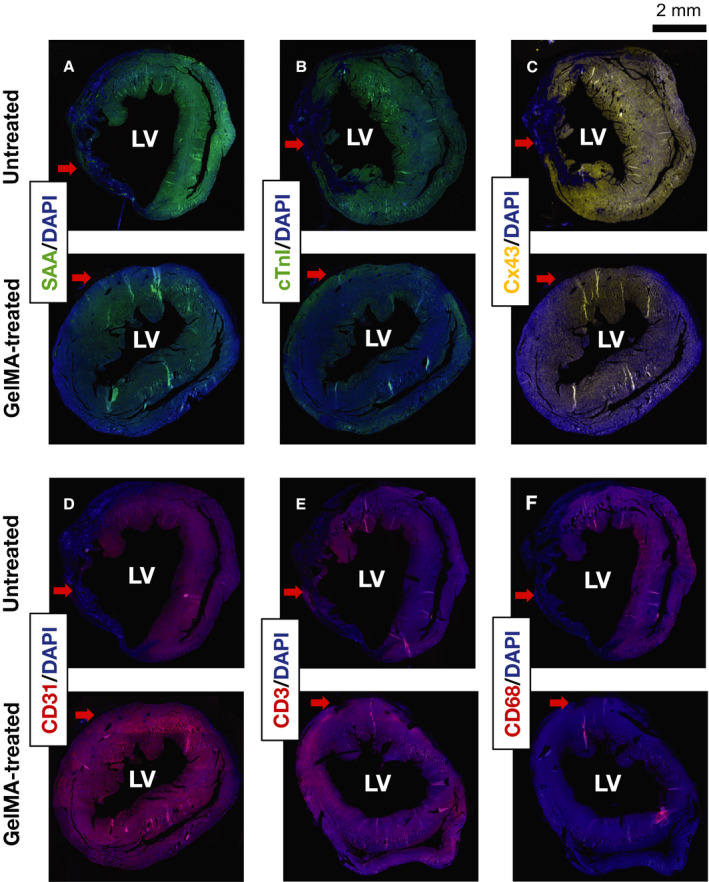
Figure 6. Immunofluorescent staining of short‐axis sections from gelatin methacryloyl (GelMA)–treated and untreated mice.

Representative images of immunofluorescent staining of untreated and GelMA‐treated hearts against sarcomeric α‐actinin (SAA) (A), cardiac troponin I (cTnI) (B), connexin 43 (Cx43) (C), CD31 (D), CD3 (E), and CD68 (F). Samples were counterstained with 4′, 6‐diamidino‐2‐phenylindole (DAPI). Red arrows indicate the area of the left ventricle (LV) affected by the ligation of the anterior interventricular artery leading to myocardial infarction.
