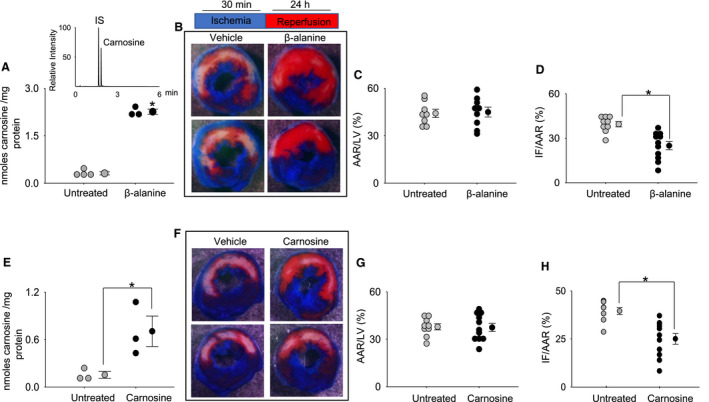
Figure 1. β‐alanine and carnosine feeding protect against ischemia reperfusion injury.

A. Wild‐type C57BL/6 mice were provided drinking water without (n=4) or with β‐alanine (20 mg/mL; n=3) for 7 days. Myocardial carnosine levels were measured by ultra‐performance liquid chromatography/tandem mass spectrometry. Inset is a representative chromatogram of carnosine and internal standard (IS) tyrosine histidine. B, Hearts of untreated and β‐alanine–fed mice subjected to 30 minutes of ischemia followed by 24 hours of reperfusion. Representative cross‐sections of 2,3,5 triphenyltetrazolium chloride staining shows the infarct zone depicted by a white area, the red area is the area at risk (AAR), and the blue region is the remote zone. (Scale bar=1 mm.) C and D, The relative ratios of the AAR to the left ventricle (LV) area and the infarct area (IF) to the AAR were compared between the untreated (n=8) and β‐alanine–fed (n=9) fed mice. E, Mice provided drinking water with or without carnosine (10 mg/mL) for 7 days; n=3 mice in each group. F, Representative cross‐sections of the untreated and carnosine‐treated mice subjected to 30 minutes of ischemia followed by 24 hours of reperfusion. G and H, Relative ratio of AAR to the LV area and IF to the AAR were compared between the untreated (n=9) and carnosine‐treated mice groups (n=11). *P<0.05 vs the untreated mice group.
