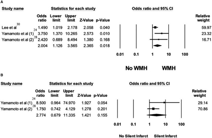
Figure 2. Forest plot of the association between reverse‐dipping pattern and silent cerebral small vessel disease neuroimaging features.

A, Forest plots of included studies assessing the association between reverse‐dipping pattern and white matter hyperintensity. B, Forest plots of the included studies assessing the association between reverse‐dipping pattern and asymptomatic lacunar infarct. A diamond data marker depicts the overall rate from included studies (square data markers) and 95% CI. WMH indicates white matter hypersensitivity.
