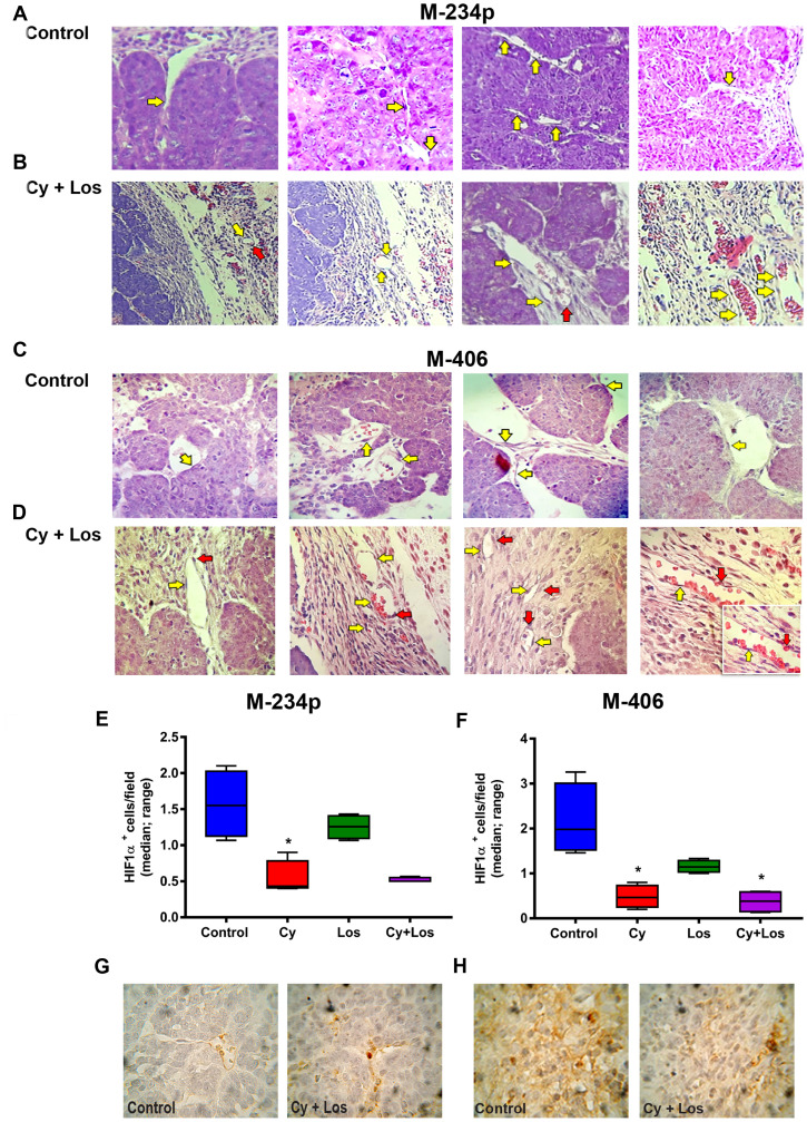
Figure 3. Vascular normalization.
Hematoxylin and eosin (H&E) representative tumor sections from M-234p and M-406, 400×. In both models the behavior was similar. Control group: (A) M-234p and (C) M-406: capillaries with small endothelial cells with barely stained nuclei and intercellular gaps (yellow arrow), lack of pericytes or cells with structure and staining compatible with pericytes. Cy+Los group: (B) M-234p and (D) M-406: intra- and peritumoral capillaries with structure and morphology similar to normal tissues. Endothelial cells with defined nuclei provide a continuous uninterrupted lining (yellow arrow), and well defined basal membrane covered with pericytes (red arrow). M-406 magnified section (1000×): vessel with normal vascular morphology. HIF1α expression: HIF1α+cells/field (median, range). (E) Control vs Cy (P < 0.05), (F) Control vs Cy (P < 0.05), vs Cy+Los (P < 0.05), (G) and H), representative images of Control and Cy+Los treated tumors, 100× magnification. Kruskal-Wallis multiple comparison test and Dunn’s post-test.

