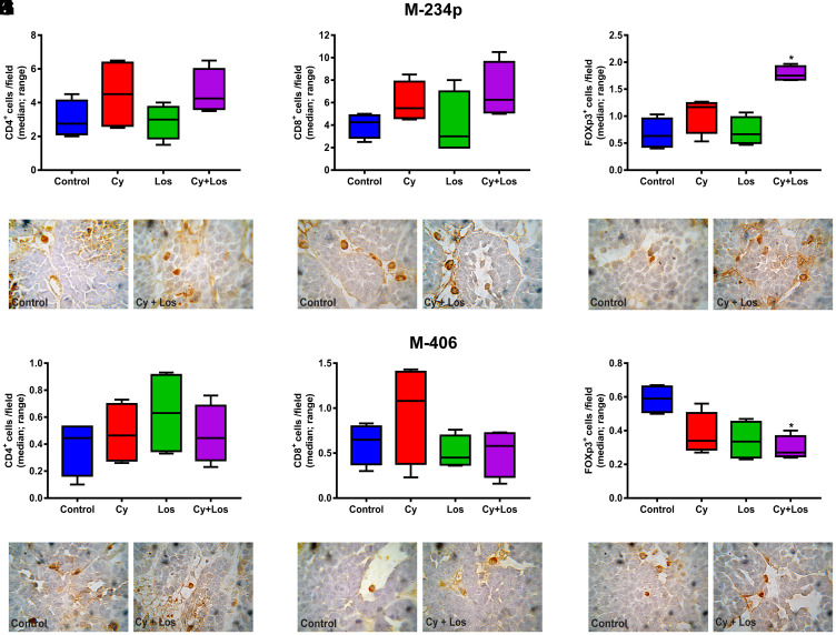
Figure 5. Quantification of tumor infiltrating lymphocytes by IHC.
Lymphocytes/field (median, range). M-234p: (A) CD4+ cells, N. S. (B) CD8+ cells, N. S. (C) Foxp3+ cells: Control vs Los, P < 0.05, vs Cy+Los, (P < 0.05); (D–F) representative images of Control and Cy+Los treated tumors, 100× magnification; M-406: (G) CD4+ cells, N. S. (H) CD8+ cells, N. S. (I) Foxp3+ cells: Control vs Cy+Los, (P < 0.05); (J–L) representative images of Control and Cy+Los treated tumors, 100× magnification. Kruskal-Wallis multiple comparison test and Dunn’s post-test.

