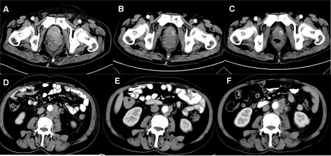
Figure 2.
Imaging material of prostate cancer. (A) Before radiotherapy combined with olaparib, computed tomography (CT) of the pelvis showed a primary prostate tumor, which invaded the bilateral seminal vesicle gland and closed to the rectum in December 2018. (B) The size of prostate tumor shrank significantly after radiotherapy combined with olaparib in January 2019. (C) Primary tumor remained stable after radiotherapy combined with olaparib in February 2019. (D) Before using olaparib, computed tomography (CT) of the pelvis showed enlarged lymph nodes adjacent to aorta abdominalis in December 2018, which was not included into the radiation field. (E) After using olaparib, lymph nodes adjacent to aorta abdominalis shrank significantly in January 2019, which was not included into the radiation field. (F) After 2.5 months with olaparib, lymph nodes adjacent to aorta abdominalis enlarged again in February 2019, which was not included into the radiation field.

