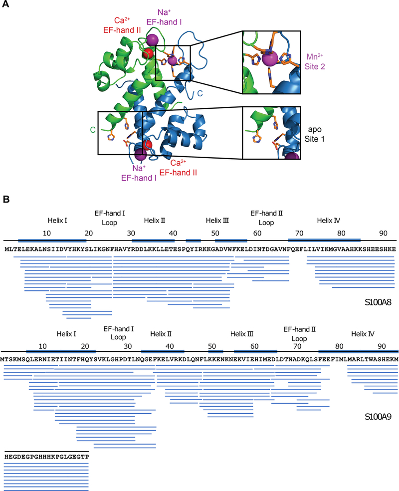
Figure 1.
hCP structure and sequence coverage map of pepsin-digested CP-Ser. (A) Crystal structure of Mn2+-, Ca2+-, and Na+-bound CP-Ser (PDB 4XJK).18 A heterodimer unit taken from the heterotetramer is shown. S100A8 is green and S100A is blue. (B) Sequence coverage map of pepsin-digested S100A8(C42S) (top), and S100A9(C3S) (bottom) of CP-Ser. Each blue bar represents a peptide identified in a tandem mass spectrometry mapping experiment. Secondary structure elements are indicated by blue lines on top of the sequence. The Ca2+-binding loops are indicated by text only.

