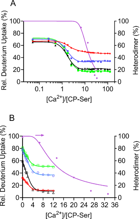
Figure 7.
Data fitted with NLLS to a model incorporating four independent Ca2+ (1–4 Ca2+) in the dimer and 8 Ca2+ in the tetramer. (A) The data are PLIMSTEX (4 μM CP-Ser) and native MS (15 μM CP-Ser) and represent the binding sites A9(C3S) 23–37 (+2) (red diamonds), A8(C42S) 27–34 (+2) (blue circles), A9(C3S) 66–78 (+2) (black triangles), A8(C42S) 55–68 (+2) (green squares), and native-MS data (purple diamonds). (B) The data are sharp-break PLIMSTEX (40 μM CP-Ser) and native-MS (15 μM CP-Ser). The data are from the Ca2+-binding sites A9(C3S) 9–13 (+1) (red diamonds), A8(C42S) 27–39 (+2) (blue circles), A9(C3S) 61–65 (+1) (black triangles), A8(C42S) 35–45 (+2) (green squares), and native-MS data (purple diamonds).

