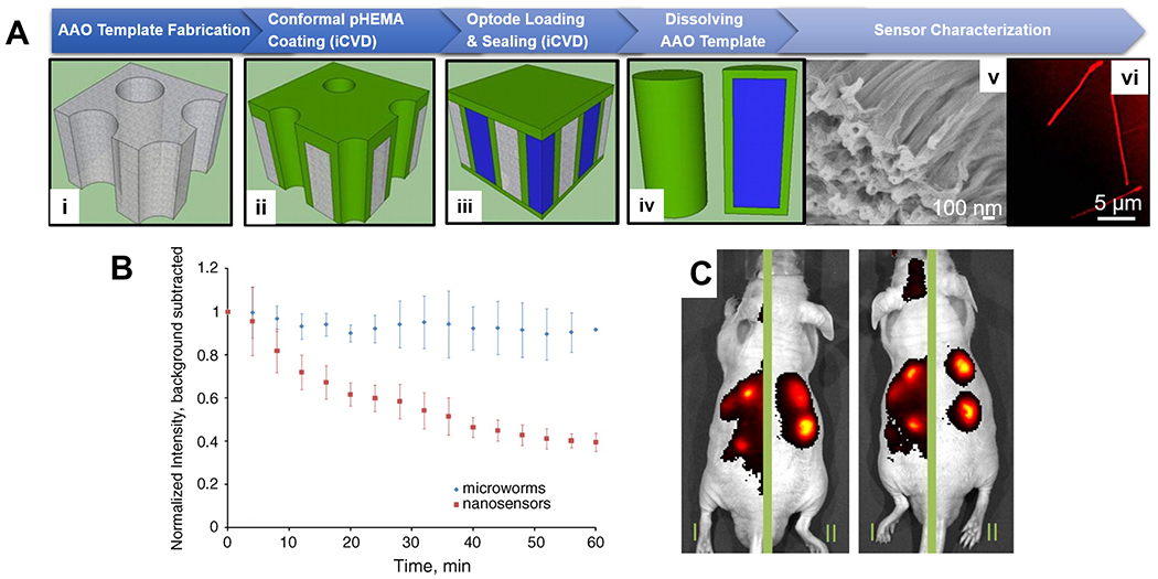
Figure 5. CVD polymer coatings for in vivo biomedical sensing.

(A) Fabrication (i-iv) and imaging (v-vi) of in vivo sodium sensors, termed microworms, enabled by the iCVD technique. Thin layers of pHEMA was deposited conformally onto AAO membranes, the pores of which were then filled with a sodium-responsive optode solution (liquid in blue) and sealed via a subsequent pHEMA deposition. Shape of the fluorophore-encapsulating microworms was confirmed with SEM and confocal microscopy. (B) Decay of the normalized fluorescent intensity over time at the site of subcutaneously injection of the in vivo sensors. The microworms (blue diamonds) exhibited longer local retention time. (C) IVIS images of animals that received regular nanosensors on the left-side of their body and the microworms on the right side, showing greater localization and retention of the microworms after injection.123
