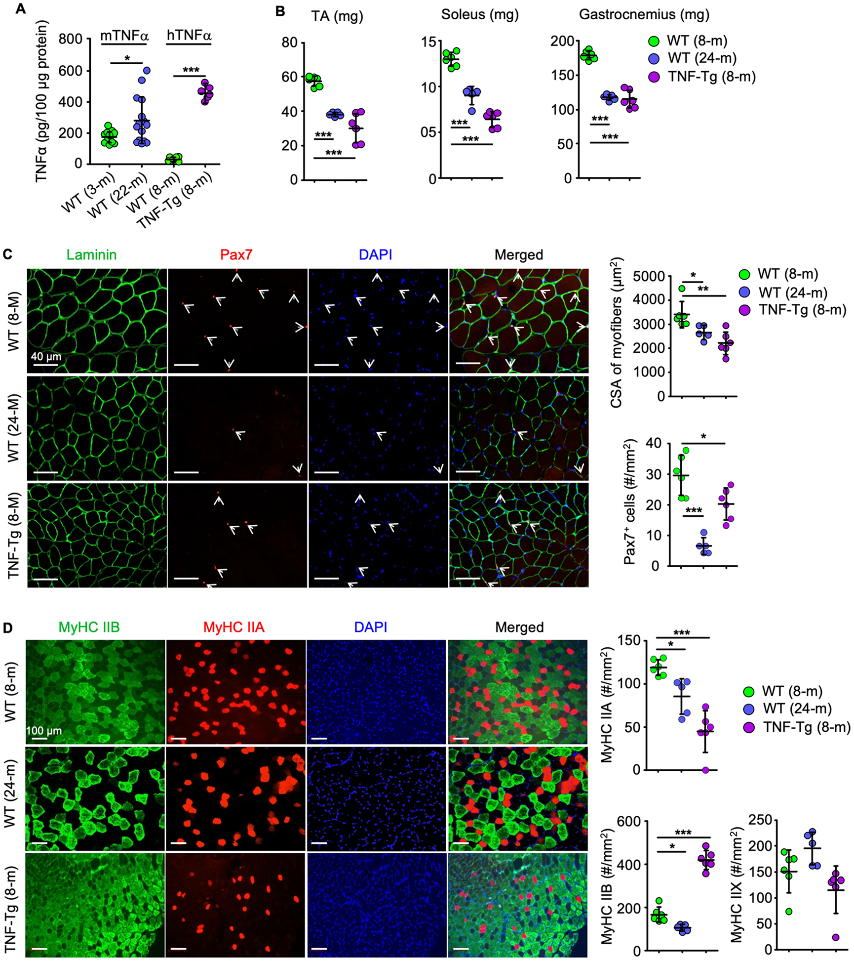
Figure 1. TNFα induces muscle atrophy in aged WT and adult TNF-transgenic mice.

(A) Murine (m) and human (h) TNFα levels in protein lysates from gastrocnemius from 14 young and 15 old male C57BL6/J mice and 6 WT and 6 TNF-Tg male mice. *p < 0.05, ***p < 0.001. (B-D) Muscle phenotypes of 6 adult and 5 old WT and 6 adult TNF-Tg mice. (B) Lean weights of tibialis anterior (TA), soleus and gastrocnemius muscles. (C) 10 μm-thick cryosections of TA muscles stained for laminin (green) and Pax7 (red) expression to assess the cross-sectional area (CSA) of myofibers and the numbers of Pax7+ satellite cells (white arrows). (D) 10 μm-thick cryosections of TA muscles IF-stained for myosin heavy chain (MyHC) IIA and IIB expression and the numbers of type IIA (red), IIB (green) and IIX (unstained) fibers. *p < 0.05, **p < 0.01, ***p < 0.001.
