
Figure 7: Endogenous Enterobacteriaceae exacerbate signs of disease in streptomycin-treated mice on a high-fat diet.
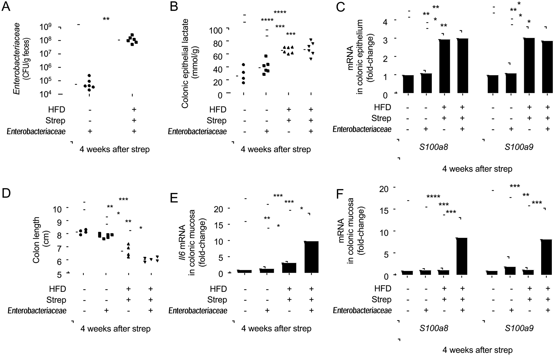
Groups of male Enterobacteriaceae-free mice (N = 6) were reared on a high-fat diet (HFD: +) or on a low-fat diet (HFD: −) and were mock treated (Strep: −) or treated with a single dose of streptomycin (Strep: +) four weeks before necropsy. One day after streptomycin treatment, mice were mock inoculated (Enterobacteriaceae: −) or received commensal isolates of E. coli, K. oxytoca and P. vulgaris (Enterobacteriaceae: +). (A) Colony-forming units (CFU) of Enterobacteriaceae in the feces were determined. (B) Lysates of colonocytes were used to measure concentrations of lactate. (C) Transcript levels of the indicated genes were determined by quantitative real-time PCR in RNA isolated from colonocyte preparations. (D) Colon length was determined during necropsy. (E and F) Transcript levels of Il6 (E), S100a8 and S100a9 (F) were determined by quantitative real-time PCR in RNA isolated from the colonic mucosa. *, P<0.05; **, P<0.01; ***, P<0.001; ****, P<0.0001. (A,C,E,F) Data were transformed logarithmically before analysis. P values were calculated by Student’s t test (A) or by one-way ANOVA followed by Tukey’s multiple-comparison tests (B-F). See also Table S2.