
Figure 2.
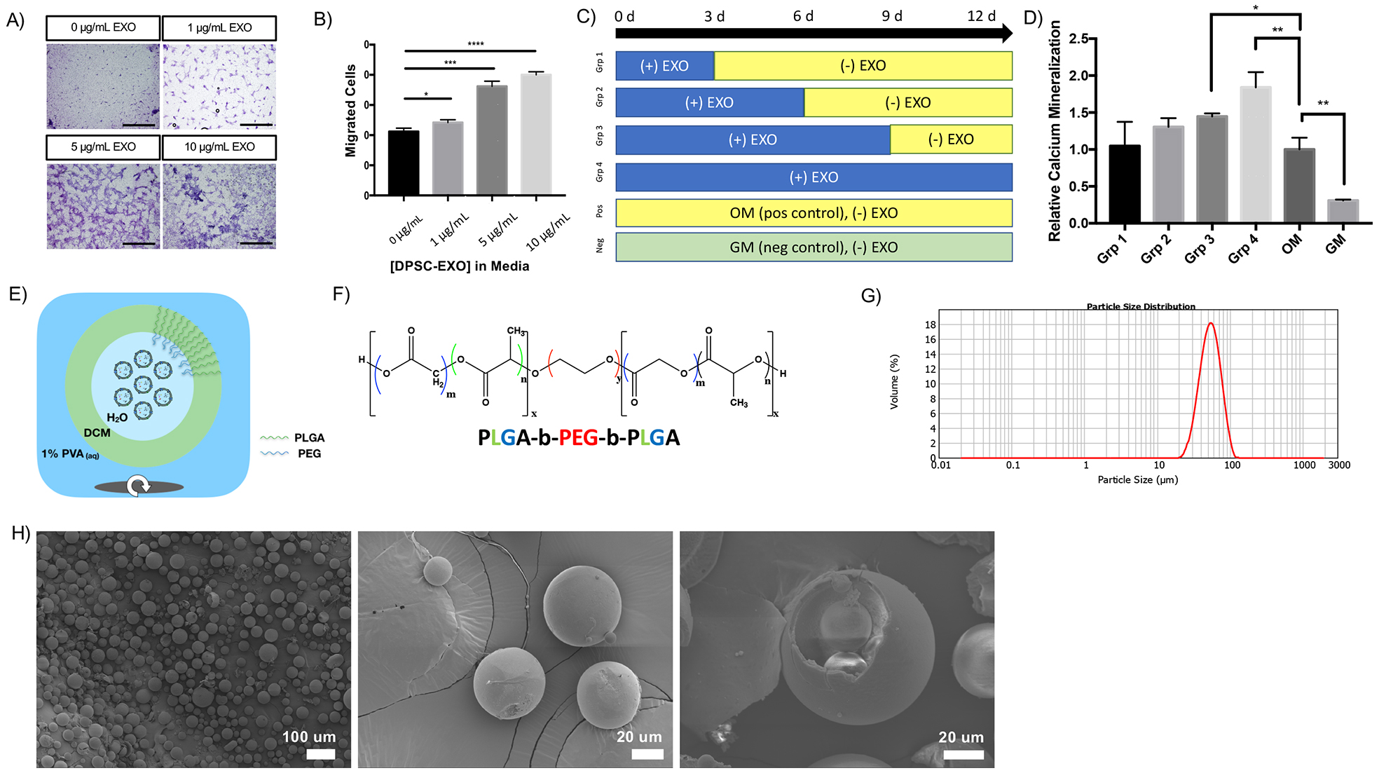
In a transwell migration assay, DPSC-EXOs are shown to facilitate migration of nascent hDPSCs in a dose-dependent manner (A, scale = 200 μm, B). Prolonged exposure to DPSC-EXO in culture media results in increased mineralization, in a time-dependent manner where the duration of exposure correlates to the degree of mineralization by DPSCs, compared to odontogenic and growth media controls (C, D). The combination of these properties lends to the development of an in vivo delivery system for dentinogenic exosomes. Exosomes are encapsulated into polymeric microspheres by a double emulsion method (E). Exosomes, concentrated in PBS, are emulsified in a dichloromethane which contains PLGA-PEG-PLGA copolymer (F). The resulting emulsion is emulsified in 1% w/v polyvinyl alcohol to form discrete water-oil-water (w/o/w) droplets which contain exosomes in their inner water compartment. After solvent evaporation, resulting particles are collected and lyophilized. Their size is measured by Mastersizer laser diffraction (G, dav g = 51.885 μm) and particle morphology is assessed by scanning electron microscopy (H – left, scale = 100 μm; middle, scale = 20 μm; right, scale = 20 μm). * p < 0.05, ** p < 0.01, *** p < 0.001, **** p < 0.0001.