
Figure 4.
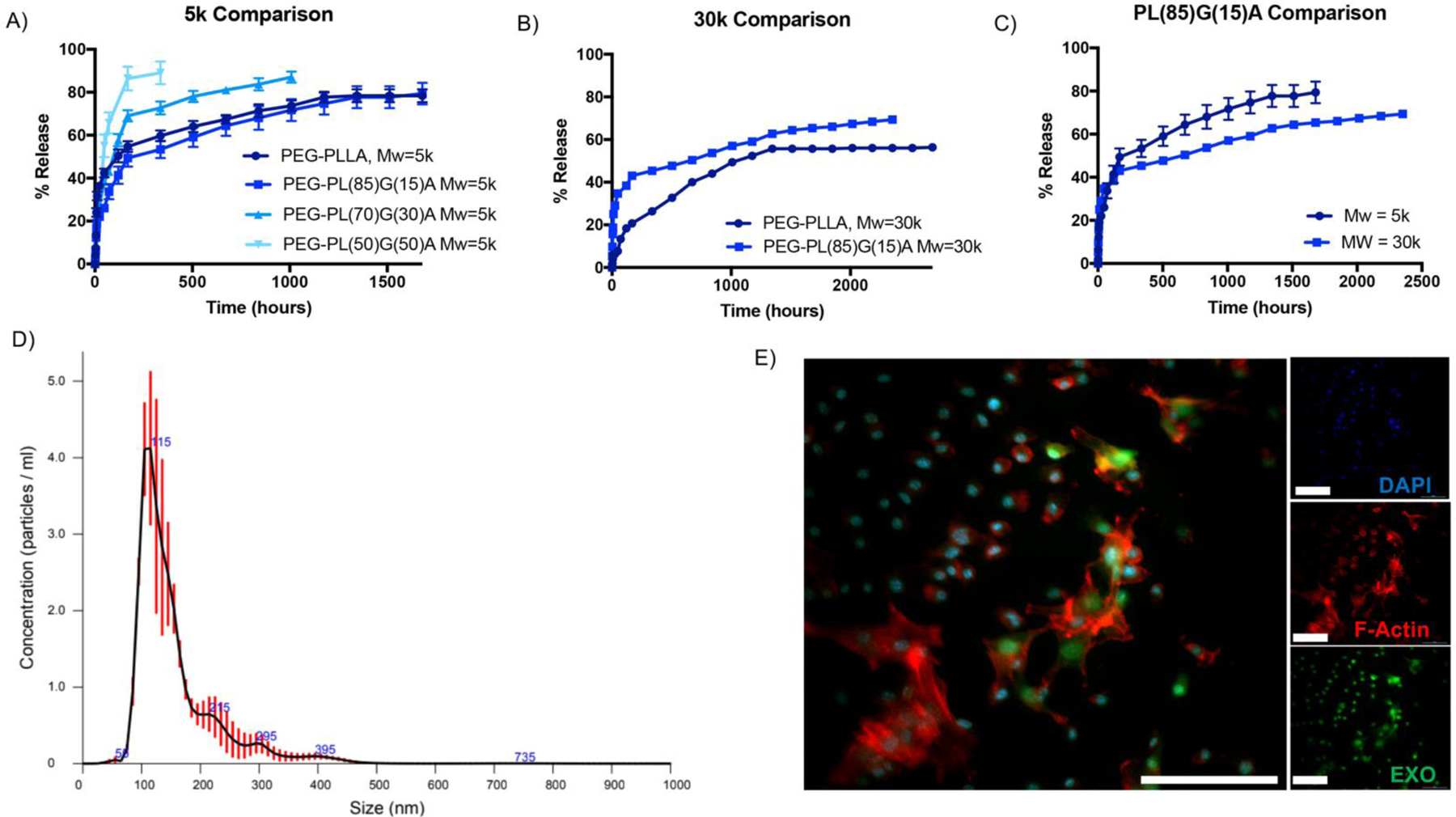
Exosomes are released from EXO-MS when incubated in vitro. The release profile is a function of molecular weight and hydrophilicity of the PLGA-PEG-PLGA copolymers (A, B, C). Released exosomes maintain their characteristic hydrodynamic diameter and size distribution at 1-week time point (D), indicating that the lipid membrane remains intact and undisrupted throughout encapsulation and release. DiO-labelled exosomes were encapsulated and released from the EXO-MS, and their rapid uptake into the cytoplasm of recipient hDPSCs was visualized by confocal microscopy (E, 30 minutes incubation. Blue = DAPI, Red = F-actin, Green = DPSC-EXO, scale = 100 μm).