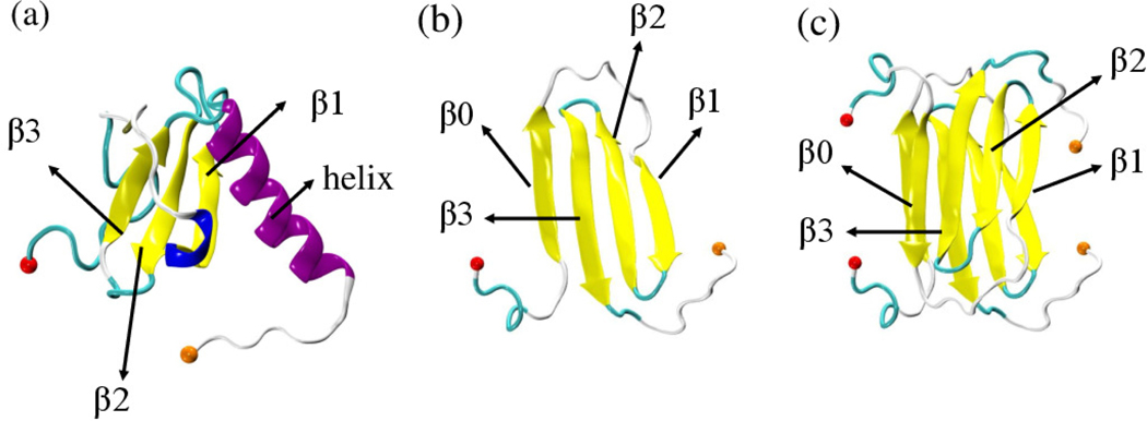
Figure 1:
Lymphotactin chains can take two distinct structures, both deposited in the Protein Data Bank: (a) Ltn10 (PDB-ID: 2HDM) and Ltn40. The Ltn40 monomer is shown in (b) and derived from the experimentally observed dimer (PDB-ID: 2JP1) shown in (c). Labels 30 identify the secondary structural elements, and the N-terminal and C-terminal Cα atoms are drawn as spheres in red and orange, respectively.

