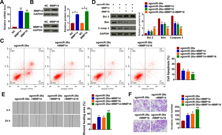
Figure 5.
miR-26a inhibits proliferation, migration and invasion by targeting MMP14 and MMP16. MMP14 or MMP16 plasmid or its NC was transfected into SCL-1 cells. qRT-PCR (A) and Western blot (B) was used to detect the transfection efficiency of MMP14 or MMP16 (*p<0.05, **p<0.01 vs NC). (C) The apoptosis of cells was calculated by flow cytometry in SCL-1 cells (*p<0.05 vs agomiR-26a, #p<0.05 vs agomiR-26a+MMP14 and agomiR-26a+MMP16). (D) Western blot was performed to detected the expression of apoptosis-related protein Bcl 2, Bax and C-casp 3 (cleaved caspase-3) (*p<0.05 vs agomiR-26a, #p<0.05 vs agomiR-26a+MMP14 and agomiR-26a+MMP16). (E) Wound healing assay was used to detect cell migration (*p<0.05 vs agomiR-26a, #p<0.05 vs agomiR-26a+MMP14 and agomiR-26a+MMP16). (F) Transwell assay was performed to check cell invasive ability (*p<0.05 vs agomiR-26a, #p<0.05 vs agomiR-26a+MMP14 and agomiR-26a+MMP16). The above measurement data were expressed as mean ± standard deviation. Data among multiple groups were analyzed by one-way ANOVA, followed by a Tukey post hoc test. The experiment was repeated in triplicate.

