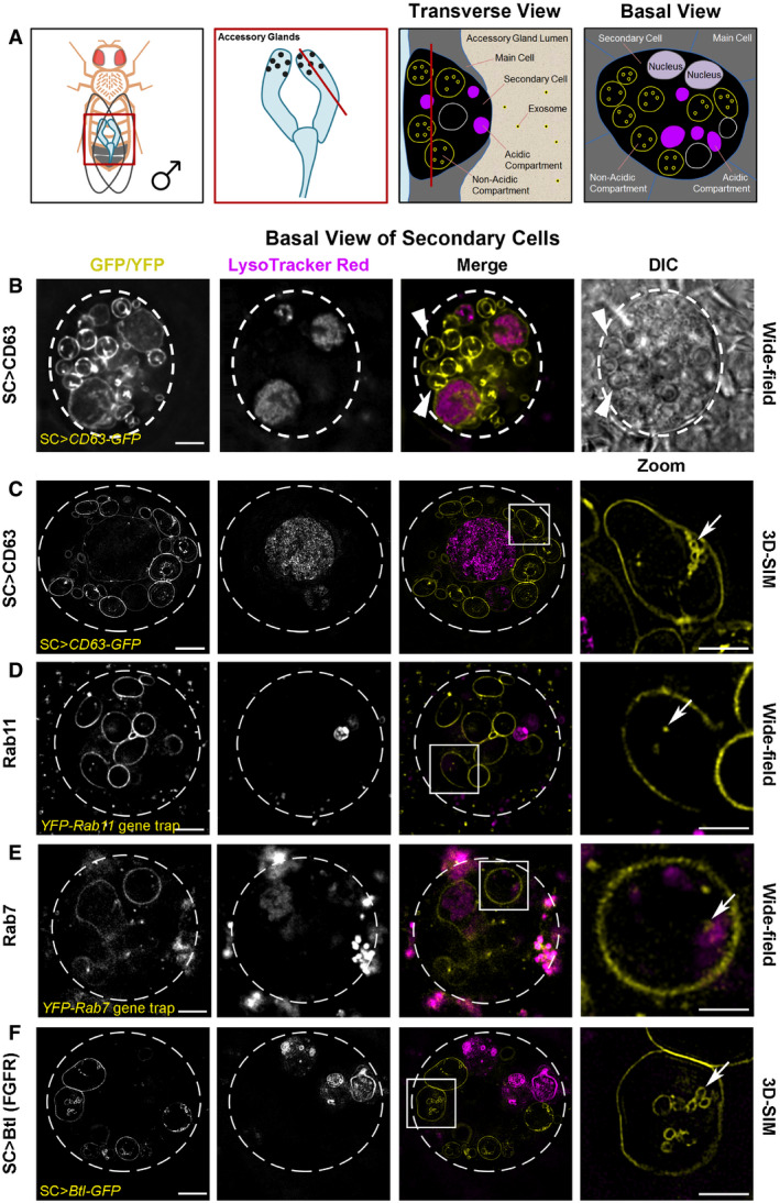
Basal views through living SCs, with dashed white circles approximating the outline of a single SC, and acidic compartments marked by the vital dye LysoTracker® Red (magenta). In merge images, a single non‐acidic (C, D and F) and acidic (E) compartment containing intraluminal vesicles (ILVs) is boxed and magnified in the right panel (Zoom). ILVs appear as membrane‐delineated vesicles, using super‐resolution 3D‐structured illumination (3D‐SIM) microscopy for the brighter overexpressed GFP‐tagged constructs (yellow; C and F). However, ILVs appear only as puncta, using lower resolution wide‐field microscopy for the fainter endogenously expressed YFP‐tagged Rab GTPases (yellow; D and E). (B) Wide‐field fluorescence image, including differential interference contrast (DIC), of SC expressing a GFP‐tagged version of human CD63 (CD63‐GFP). CD63‐GFP expression is apparent on the limiting membranes of non‐acidic compartments and their ILVs and also on the limiting membranes of the enlarged acidic compartments. Most large non‐acidic compartments are Rab11‐positive (D) and contain dense‐core granules, which have a “fried egg” appearance with DIC (arrowheads) (Corrigan
et al,
2014; Redhai
et al,
2016). (C) 3D‐SIM image of CD63‐GFP-expressing SC. Arrow highlights CD63‐GFP-marked ILVs (Zoom). Many more ILVs are apparent in non‐acidic compartments in a complete Z‐stack of a non‐acidic compartment (
Movie EV1). (D) Wide‐field fluorescence image of an SC expressing a
YFP‐Rab11 gene trap. YFP‐Rab11 marks the limiting membranes of most non‐acidic compartments and internal puncta (arrow in Zoom), but not the surface of acidic compartments (
Appendix Fig S1B). (E) Wide‐field fluorescence image of SC expressing a
YFP‐Rab7 gene trap. YFP‐Rab7 marks the limiting membranes of acidic compartments and internal puncta (arrow in Zoom). Enlarged acidic compartments are also present in adjacent main cells. (F) 3D‐SIM image of SC expressing a GFP‐tagged version of Breathless (Btl‐GFP). Btl‐GFP marks the limiting membranes of non‐acidic compartments and their ILVs (arrow in Zoom), but not the surface of acidic compartments (
Appendix Fig S1C). Images from 6‐day-old male flies shifted to 29°C at eclosion. This induces GAL4/UAS-dependent SC transgene expression in (B, C and F). The genotypes of flies carrying multiple transgenes are as follows:
w; P[w
+
, UAS‐CD63-GFP] P[w
+
, tub‐GAL80
ts
]/
+;
dsx‐GAL4/
+ (B and C);
w; P[w
+
, tub‐GAL80
ts
]/
+;
dsx‐GAL4/
P[w
+
, UAS‐btl-GFP] (F).

