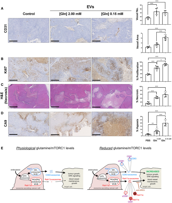
HCT116 flank tumours produced by subcutaneous injection were established for 20 days before injection at 3‐day intervals with vehicle (PBS), or EVs isolated by UC from glutamine‐replete or glutamine‐depleted HCT116 cells. Tumours were excised 24 h after last of four injections for analysis. Panels show representative immunostained histological sections of tumour tissue quantified using the Visiopharm Integrator System.
Sections immunostained for CD31, which labels endothelial cells and blood vessels, with blood vessel number (upper) and total area (lower) represented in bar charts.
Sections immunostained for Ki67, which stains proliferative cells. Proportion of tumour cells with Ki69 staining is represented in bar chart.
Sections stained with haematoxylin and eosin, which highlights necrotic regions (pale staining). Proportion of tumour area that is necrotic is represented in bar chart.
Sections immunostained for CA9, which is expressed in hypoxic regions. Proportion of tumour cells with CA9 staining is represented in bar chart.
Schematic model showing how in cancer cells, regulation of endosomal trafficking by depletion of exogenous glutamine or reduced Akt/mTORC1 signalling can induce a change in the balance of exosome production. Relative levels of a mixed population of exosomes from the established Rab7‐late endosomal multivesicular bodies (MVBs), termed “CD63‐exosomes”, are reduced relative to the mixed exosome population from Rab11a‐labelled recycling endosomal MVBs, termed “Rab11a‐exosomes”, a subset of which appear to be marked with Rab11a. Although CD63 does not appear to be trafficked through these latter compartments, the “classical” exosome marker CD81 appears to be present at undefined levels and marks some Rab11a‐containing exosomes. The resulting vesicles can increase ERK signalling and cell growth in recipient tumour cells and enhance growth and stability of network formation in endothelial cells. The growth effects are Rab11a‐dependent and can be blocked by anti‐AREG antibodies. Thickness of arrows indicates relative levels of membrane flux through the two exosome‐generating endosomal routes.
Data information: Scale bar is 250 μm, except for (C), which is 500 μm. Data were analysed by one‐way ANOVA;
n ≥ 4; *
P < 0.05, **
P < 0.01, ***
P < 0.001. Bars and error bars denote mean ± SD.
Source data are available online for this figure.

