FIGURE 3.

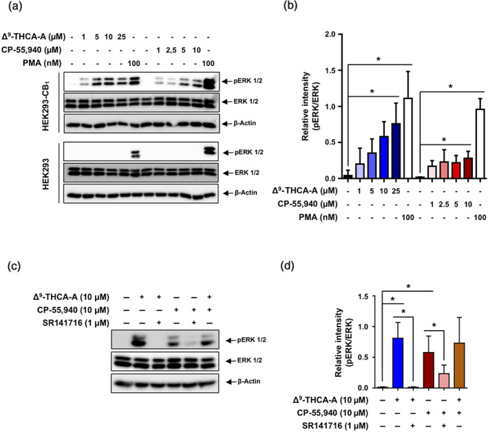
Effects of Δ9‐THCA‐A on ERK1/2 phosphorylation. (a) Western blot analysis showing p‐ERK1/2, total ERK1/2, and actin levels in HEK293‐CB1‐β‐arrestin Nomad cells and HEK293 cells exposed to increased concentrations of Δ9‐THCA‐A and CP‐55,940 for 30 min. (b) Quantification of western blot results from five independent experiments. (c) Western blot analysis in HEK293‐CB1‐β‐arrestin Nomad cells pre‐stimulated with Δ9‐THCA‐A (10 μM) and SR141716 (1 μM) for 5 min and treated with CP‐55,940 (10 μM) for 30 min. (d) Quantification of western blot results from five independent experiments. PMA (100 nM) was used as a positive control of ERK1/2 phosphorylation. Data are means ± SD; n = 5. * P < .05, significantly different as indicated
