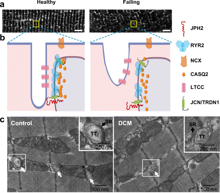
Fig. 2.
Altered dyad architecture in heart failure. (A) Confocal micrographs of the T-tubule system revealed by MM4-64 membrane dye in healthy and diseased murine cardiomyocytes. Note the T-tubular disorganization in the diseased cardiomyocyte. (B) Schematic illustration of normal (left) and diseased (right) dyadic junctions. The diseased dyad architecture features T-tubule loss and remodeling, ion channel dislocation, junctional contact area shrinkage, LTCC-RYR2 uncoupling, RYR2 cluster redistribution, and disordered fuzzy space (see text). Bar = 5 μm. JCN, junctin; TRDN1, triadin 1. (C) Transmission electron micrographs of human LV myocardium from healthy control (left) or patient with dilated cardiomyopathy (DCM, right).Arrows point to T-tubules (TT). Boxed areas are enlarged in upper right corner. In control, jSR is separated from T-tubule by a narrow junctional gap. In DCM, the junctional gap is wider, and the contact area is reduced. Panel C was reproduced from Zhang et al. (2013) with permission from the publisher

