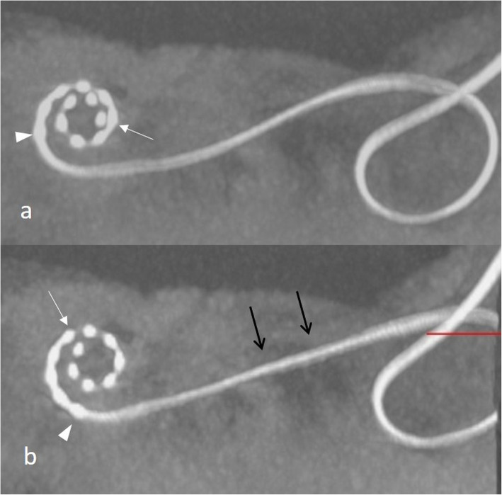
Fig. 34.
A 53-year-old male patient, with a complete malfunction of the CI and pain 3 months after CI surgery. CBCT paraaxial maximum intensity projection image shows initial overinsertion of the electrode array (a). The control scan shows back extrusion of the electrode array with migrated positions of the electrode tip (arrow) and basal electrode element (arrowhead) and straightened electrode array (black arrows) in the mastoidectomy cave (b)

